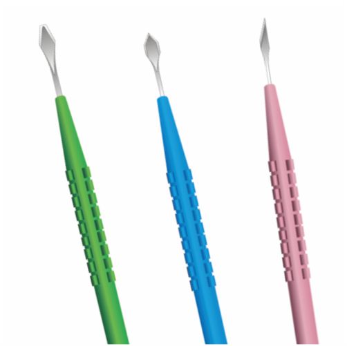
Intraocular inflammation is caused by retention of foreign material like fibers after intraocular surgery or from introduction of starch into the eye at the time of surgery. Debris can enter the eye if the doctor is unaware of its presence. The denaturation on ophthalmic equipment may be caused by the disinfection 1, 2 3 and sterilization process. The exact nature of the material that is deposited on the surfaces remains unclear.
It may be possible that it is the viscoelastic substance to which the introducer is exposed, during the insertion of the IOL into the capsular bag. Drying of the viscoelastic material may have made it difficult to remove during the routine sterilization process. Microscopic inspection of instruments after decontamination has inadequately processed instruments. Loose debris like the fibers on the tips of these tools could have been attracted to them by static forces.
Also, the fiber on the optic of the IOL could have been attached to it from the surface it was laid on at the time of folding. Foldable lenses should be laid on fiber-free surfaces. The sheets used to cover the surface should be made of fiber-free material and the equipment should only rest on surfaces that do not have such fibers. Deposits are predominantly found on the surfaces of the forceps that grasp the foldable IOL.
It is important to inspect the equipment under the operating microscope to identify the presence of debris. Once identified, the loose debris like the 4 5 6 fibers can be removed before using the tool. Clean equipment from another tray should be used if deposits are found. The silicone material can allow adhesion of bacteria and other biomaterials. The foreign material from any instrument can get deposited on the surface of the IOL and gain entry into the eye.
CAUSE OF DEBRIS ON OPHTHALMIC DEVICES
Debris can be caused by: Inadequate cleaning, rinsing and drying, using tap water, and malfunctioning autoclave. Most equipments are made from stainless steel. It does not usually rust. However, it can corrode if it is left to soak for a long period of time in any liquid. Once the tool has started to rust it will become weak, and the rust will eventually destroy and break the equipment. Rust mostly occurs on chrome or nickel-plated devices.
When the plating wears off, the steel is exposed and is further corroded by washing. If this occurs the equipment’s cannot be sterilized properly. Inexpensive Ophthalmic instruments and equipments tend to rust more easily as the stainless steel is of low quality. Sodium nitrate is an anti-rusting agent and can be used in conjunction with other the lubricants. Tablets can be dissolved in 500ml of water, when washing these devices.
Instrument stains via thorough inspection can reveal discoloration of the metal. Some stains may be rubbed off with a rubber eraser but it may leave a rough surface. Contact with hydrochloric acid and iodine must be avoided. Instruments not rinsed thoroughly after chemical sterilization can impact stain. Manufacturer’s recommended soaking times should not be exceeded.
ROLE OF CLEANING AT THE END OF THE PROCEDURES
Cleaning distilled water is preferred. If neither is available, freshly boiled tap water can be used. The following method should be used after each procedure. Three containers are required: Container I: hot soapy water. The instrument should be supported carefully whilst cleaning. A soft toothbrush may be used to clean each device individually. Needle holders, scissors, as well as artery forceps must be completely opened to clean inside the jaws.
Cannulae should be flushed through. Lens matter and visco-elastic gel block cannulae permanently. Cotton wool must not be used to clean instruments as it damages tips as well. Container II: lubricant prevents the development of stiff joints and limits the development of corrosion. Open in a separate window, ophthalmic tool need particularly careful handling. Photo: Kikuyu Eye Unit Lubricant is also needed for hinged instruments only like scissors, needle holders, as well as artery forceps. The instruments are usually dipped, one by one, into the lubricant; they should not be soaked If a lubricant is not available, the medical equipment should be rinsed in clean hot water.
One should not put cannulae in lubricant. Container III: clean hot water, as well as excess lubricant or soap is rinsed off and the equipment is left in the open, dismantled position on a clean absorbent cloth. Cannulae must be flushed via again to remove any soap debris. Drying Instruments must be dried completely before being stored. If the instruments are put away wet, they will rust. Hair dryer is very effective for drying the joints of instruments. If it is not available, dry gauze may be used cautiously. Never force open a forceps even while drying, as this will distort the instrument.
CONCLUSION
Thus, processed ophthalmology instruments have debris on them that may gain entry into the eyes at the time of surgery. This may be responsible for intraocular inflammation and other infections as well. These instruments can also pose a risk of transmission of infective agents of other diseases. Every possible measure must be undertaken to ensure the cleanliness of the instruments before using it. The measures that are discussed can help in preventing the deposits from occurring.
The answer may lie in processing ophthalmic tools via routine ultrasonic cleaning and using a chemical process to remove the debris, well before the sterilization. Simple measures like mechanical cleaning, especially those that are prone to develop these deposits, in the operating theatre can help to avoid this problem. The surgeon should still be vigilant and inspect instruments under the operating microscope before any sort of use.
